EYE HEALTH
At Kiddies Eye Care, we utilise a number of technologies and equipment to comprehensively assess your or your child’s eye health. These tests include, but are not limited to:
retinal imaging and oct
Optical Coherence Tomography
Optical coherence tomography (OCT) is a non-invasive imaging test.
Similar to an ultrasound, it uses light waves to take cross-section pictures of your retina.
With an OCT, your optometrist can see each of the retina's distinctive layers and predict early nerve loss.
Fundus Photography: Retinal Imaging
Fundus Photography/Retinal imaging takes a digital 2D flat picture of the back of your eye.
It shows the retina, the optic disc and blood vessels.
dry eye clinic
Dry eye is a common and uncomfortable occurence. Just recently, we have established a Dry Eye Clinic where we aid to alleviate the symptoms of watery and itchy eyes. In addition to providing a number of at-home options for you to bui, including Bruder Masks, specialised drops and cleaning wipes, we are also using the new Blephadex technology to irritate ectoparasitic Demodex mites in your eyelashes to resolve symptoms.
icare tonometry
Tonometers are used to measure intraocular pressure - that is, the fluid pressure inside the eye. Our technology can also be found at paediatric hospital clinics as it provides a quick and easy way to do so without any anaesthesia or air. Measuring ocular pressure has traditionally been a very uncomfortable process for most patients, however this technology assists in making it particularly more comfortable for all patients - children and babies especially.
We also have the traditional Perkins tonometers on hand.
medmont visual fields
The Medmont Visual Fields Machine can assess the visual fields of each eye separately, or also perform a binocular test to assess fitness to drive in our adult patients.
A visual field test is a subjective measure of central and peripheral vision, or “side vision,” and is used by our optometrists to diagnose any losses and their the severity, as well as monitor your glaucoma.
video slit lamp
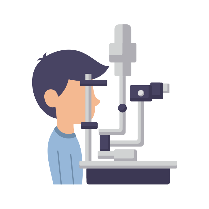
A slit lamp is a microscope that allows for assessing the anterior eye.
The optometrist can look at anterior structures such as:
lashes,
conjunctiva,
sclera,
cornea,
anterior chamber,
iris,
lens,
and vitreous
These can all be reviewed while the patient and their families can watch on the monitor in the room. Any findings will be clearly shown and explained by our optometrists.


